The Human Eye


The Human EyeThe eye is a slightly asymmetrical globe, about an inch in diameter. The front part of the eye (the part you see in the mirror) includes:

  • The iris (the pigmented part)
  • The cornea (a clear dome over the iris)
  • The pupil (the black circular opening in the iris that lets light in)
  • The sclera (the white part)
  • The conjunctiva (an invisible, clear layer of tissue covering the front of the eye, except the cornea)

Just behind the iris and pupil lies the lens, which helps to focus light on the back of the eye. Most of the eye is filled with a clear gel called the vitreous. Light projects through the pupil and the lens to the back of the eye. The inside lining of the eye is covered by special light-sensing cells that are collectively called the retina. The retina converts light into electrical impulses. Behind the eye, the optic nerve carries these impulses to the brain. The macula is a small sensitive area within the retina that gives central vision. It is located in the center of the retina and contains the fovea, a small depression or pit at the center of the macula that gives the clearest vision.

Eye color is created by the amount and type of pigment in the iris.


Diagnostic Tests

Optical coherence tomography (OCT)
OCT uses light rays to scan and image the back of the eye. This enables the fine layers of the retina to be studied. It is particularly useful to diagnose and monitor diseases of the Macula such as macula holes, macula oedema, macular degeneration and vitreomacular traction syndrome. It is also used to monitor Glaucoma as it objectively measures the thickness of the nerve fibres around the optic nerve (the principal structure damaged in poorly controlled glaucoma).


Digital photography
Digital photography of the retina are taken at the first visit and at regular intervals which allow the Ophthalmologist to document and monitor changes of the retina over time. With digital photos, the patients are able to see what their retinas look like, which helps to gain a better understanding into what the Ophthalmologist looks at during the examination.

Optical Biometry
Optical Biometry (A Scan) allows accurate calculation of the intraocular lens power required for cataract surgery. An AScan is performed in order to measure the length, curvature and power of the eye. These measurements are then used to calculate very accurately the power of the artificial lens (IOL) that will be used to replace the cataract.

Visual field testing
Visual field testing is performed to assess patients peripheral (side) vision. This is done in the clinic and is an automated test that takes approximately 20 minutes. Each eye is tested individually, and the testing process involves fixating on a central target while using your peripheral vision to react to light stimuli being projected onto various areas in your field of vision.

Visual field testing is most commonly used to assess glaucoma; however it can also be used to assess damage to the optic nerve or neural pathways from other causes such as brain tumours and strokes.

Retinal angiography
Retinal angiography fine retinal and choroidal vessels are studied using specialized digital cameras and scanning laser technology. This testing is done when the Ophthalomogist suspects a retinal disorder, particularly those that involve problems with the circulation of blood vessels in the eye. This testing provides us with more detailed assessment in retinal disorders including diabetic retinopathy, macular degeneration, and other retinal vascular diseases. Fluorescein dye is most often used for retinal angiography

Amsler grid
An Amsler grid is a simple take home test to monitor any changes in your vision. The Amsler grid has dark horizontal and vertical lines forming a grid. If any changes are detected on the grid, contact your Ophthalmologist as soon as possible. These changes include wavy lines, missing areas, or holes on the grid. The earlier the condition is detected, the higher success rate of treatment there is.

Cataract Surgery

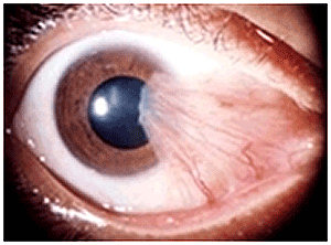
Cataract surgery is the removal of the natural lens of the eye (also called "crystalline lens") that has developed an opacification, which is referred to as a cataract. Metabolic changes of the crystalline lens fibers due to age or trauma lead to the development of the cataract and loss of transparency, causing impairment or loss of vision. Symptoms include: difficulty seeing afar, difficulty reading print, glare, frequent changes to the spectacle prescription, diminishment of colour and brightness. During cataract surgery, a patient's cloudy natural lens is removed and replaced with a synthetic lens to restore clear vision.

If a cataract is suspected or diagnosed, a complete medical eye exam will determine if the cataract needs to be removed. Specifically the clinician will perform Ultrasound size measurements of the eye to determine the most appropriate power for the replacement lens. This intraocular lens (IOL) will reduce the need for glasses and if glasses are required, they will be much thinner and lighter than if an IOL was not implanted.
The primary choices of IOLs are monofocal, multifocal, toric and accommodating. Each has its own advantages and disadvantages. Only after careful discussion with the doctor can the appropriate type of IOL can be selected.


Following surgical removal of the natural lens, an artificial intraocular lens implant is inserted. Cataract surgery is performed by an ophthalmologist in a day surgery or hospital, using local anaesthesia (either topical, peribulbar, or retrobulbar), usually causing little or no discomfort to the patient.

Phacoemusification(Phaco) is the preferred method of removal in almost all cataract operations performed today. It involves a very small incision made in eye and the use of a machine with an ultrasonic handpiece equipped with a titanium or steel tip. The tip vibrates at ultrasonic frequency (40,000 Hz) and the lens material is emulsified.

A second fine instrument (sometimes called a cracker or chopper) may be used from a side port to facilitate cracking or chopping of the nucleus into smaller pieces. Fragmentation into smaller pieces makes emulsification easier, as well as the aspiration of cortical material (soft part of the lens around the nucleus). After phacoemulsification of the lens nucleus and cortical material is completed, a dual irrigation-aspiration (I-A) probe or a bimanual I-A system is used to aspirate out the remaining peripheral cortical material.
The surgery site is then cleaned and prepared for the insertion of the artificial lens. The lens is folded and inserted using a injector and positioned into place.The small incisions do not need stitching and the wound will self seal within minutes of the operation.

The absence of stitches means the visual recovery is fast and there is little discomfort after the surgery. A protective sheild will be positioned over the patient's eye. The complete surgery process will take between 20-40 minutes.

The patient can go home on the same day as the surgery. Transportation home from the surgery needs to be arranged. The patient will be required to use both an antibiotic and steroid drop for approximately 4 weeks following surgery. Recovery is often short within 2 to 6 weeks and complete healing at three months. Some people have noticeable improvement in their vision from the day after surgery onward, while others need more time for their vision to clear.
There is no major restrictions following surgery, however it is advisable not to go swimming for 2 weeks. 

Laser cataract surgery is the latest advance.

Here, a femtosecond laser prepares the eye for phacoemusification by making all the wounds, the opening into the front part of the cataract and starts to break up the cataract into pieces. The rest of the operation is then completed as above. Its advocacy is couched in terms of greater accuracy and safety.


YAG laser capsulotomy

A small number of patients may experience capsule thickening months after cataract surgery but more commonly it occurs two or three years after. When the capsule thickens it becomes opaque. This stops the light reaching the back of the eye, as a result vision can be affected and sight becomes cloudy or misty, this will be most obvious at night, or in sunlight.
 Laser treatment makes a small hole in the centre of the capsule, and can restore vision to how it was after the cataract operation. This procedure is painless.

The procedure usually only takes a few minutes. Following a YAG laser capsulotomy, normal activities can be continued. The experience of some floaters afterward is normal and these will likely resolve within a few weeks. Most people can expect their vision to improve within a day. 
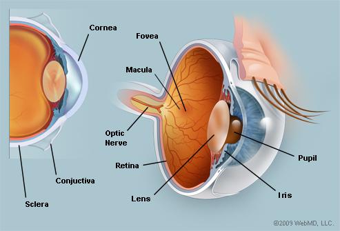
 

Minor Operations

Pterygium removal

A Pterygium is caused by UV damage to the conjunctiva which is the outer layer of skin covering the eye. An abnormal growth grows slowly over the white of the eye until it reaches the cornea. Once it starts to grow over the cornea it can cause irritation and visual distrubances. It is at this stage that removal is required.

Pterygium

In ptergium surgery, the abnormal tissue is removed from the cornea and sclera (white of the eye). A graft of conjunctiva is harvested from the eye and placed in the area from where the pterygium was removed. This surgery is performed under local anaesthetic in the clinic's treatment room or in day surgery. The procedure takes around 45 minutes and the healing process takes between 2 to 4 weeks with slight discomfort.


Eyelid Cyst

The process of removing an eyelid cyst is performed in the rooms in our fully equipped procedure room. The operation is performed under local anaesthetic, which is applied with a small injection into the eyelid. 
The chalazion (cyst) is removed from a small incision on the back of the eyelid, so it is not seen from the outer eyelid. The eyelid is lifted and inverted, a small cut (about 3mm) is made just on top of the chalazion. The lump is then removed and pressure is applied to the eye. Recovery time is minimal and no with no scarring.

Eyelid cyst

Probing and syringing

Probing and syringing is usually a quick procedure where a fine probe is passed through the tear duct to clear the blockage or to widen the tear duct. Sometimes a dye is then passed through to check that the duct is clear. This procedure is performed for patients who experience persistent watery eyes and is carried out in office.

Removal of lid lesions

Often lesions grow on the lid or lid margins that are quite safe to leave untreated but for cosmetic or visual reasons they are removed. The lid is anaesthetised using local anesthetic and a small wedge section is made in the lid, the lesion removed and the skin is sutured if required (depending on the size of the lesion).

Biopsy 
If a lesion or lump is seen on the lid or lid margin a biopsy may be necessary to determine the cause. A small section or the complete lesion is removed and sent to an external pathology company for full analysis. The results are usually received in 3 to 4 days and you will be contacted with the result.